 German
 Ελληνικά
In many cases the tears’ drainage is not completely open at birth. In fact this phenomenon is so common that the obstruction in the first months of life is almost considered normal.
Obstruction of the lacrimal drainage
In many cases the tears’ drainage is not completely open at birth. In fact this phenomenon is so common that the obstruction in the first
months of life is almost considered normal. The barrier is usually a thin film at the end of the lacrimal route, just prior to its extrusion to the nose, but may be located at any height. The obstruction results in tears not drained but poured from the eye to the child's cheeks causing discomfort and irritation.
The stagnant of tears in the eyes makes it difficult for vision, but the normal development does not appear to be influenced. Even though tears seem flowable, they also contain a quantity of mucus.
If their watery element is evaporated, mucus remains on the lashes and the child's eyelids stick together, especially in the morning. In this case, tears and mucus favor microbial growth.
Such infections occur by swelling and redness of the eyelids, but are rarely serious. Tearing seems to get worse if the child has a cold. The swelling of the tissues around the nose, which accompanies a cold, causes additional pressure and obstruction of the lacrimal drainage, resulting in deterioration of the lacrimation.
Dacryocystitis
The infection of the lacrimal sac (the wide part of the ipsilateral drain to the root of the nose) is called dacryocystitis and is manifested by intense tearing, eye gum, swelling, redness and tenderness in the area.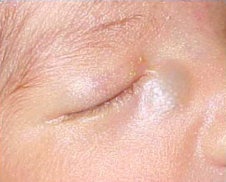
It can occur at any age, but infants are more susceptible because the development of the lacrimal system is not yet complete. The treatment consists of a gentle massage to the area between the eye and nose, to assist the opening of the pore alongside antibiotic drops or ointments.
Sometimes, at birth or a few days after, a bluish "inflating" is visible from one side of the root of the nose. This is the lacrimal sac, filled with mucus due to the obstruction of the nasolacrimal duct. This condition predisposes to the development of dacryocystitis, but may have other adverse consequences, such as difficulty in the infants’ breathing. In such cases surgery is nessesary.
Treating obstruction of the lacrimal drainage
Addressing an obstruction in the tear drainage depends on the child's age and severity of symptoms. Generally, infancy blockages have a tendency to improve; so many ophthalmologists prefer to wait before proceeding to surgical interventions.
Massaging the lacrimal sac: This technique is very simple and can be applied at home by parents. Firstly, we press gently with our index the area between the eye and the root of the nose and then we do gentle circular movements (massage) for several seconds. This is repeated at least 2 times in 24 hours. When executing the technique, the material trapped in the lacrimal sac may come from the punctum, and thus thus be carefully wiped.
Sometimes massaging the area can even break the film which caused the obstruction, and the problem will be solved immediately.
Antibiotics: Antibiotic eye drops and ointments are used to control the infection and to reduce the production of mucus, but not to cure for the actual blockage. There is an indication of prescription only when the eyelids are very red and swollen or when the secretions are many. Otherwise they should not be prescribed, since the same antibiotics can cause irritation.
Catheterization of the lacrimal ducts: It is a simple and effective process. In young infants, it can be done in the doctors’ office without anesthesia, but in older children anesthesia and surgical conditions are required.
A special small stent is inserted from the lacrimal punctum to the lacrimal duct resource, in order to break the film which caused the obstruction. The tip of the catheter is blunt and in no case do we pierce or cut the child’s skin. After the opening of the obstruction a colored liquid is administered from the lacrimal punctum and is sucked from the nose to confirm patency of the lacrimal system.
The whole process lasts a few minutes and the complications are very rare. The child is ready to return home when he wakes up from the anesthesia.
The success rates are high, but for various reasons, a 10% may need repetition or some additional surgical intervention in the nasal cavity. Usually the cases that fail are children who are catheterized a lot after completing their first year of life. If after 2-3 catheterizations the obstruction is not solved special small silicone tubes may be placed in the lacrimal duct for a few months. Usually, even after removal the pore remains open. The operation is called dakryoaskorrinostomia and is accessed through the nose with a special endoscope. Luckily, it is extremely rare children needing this surgery.