21


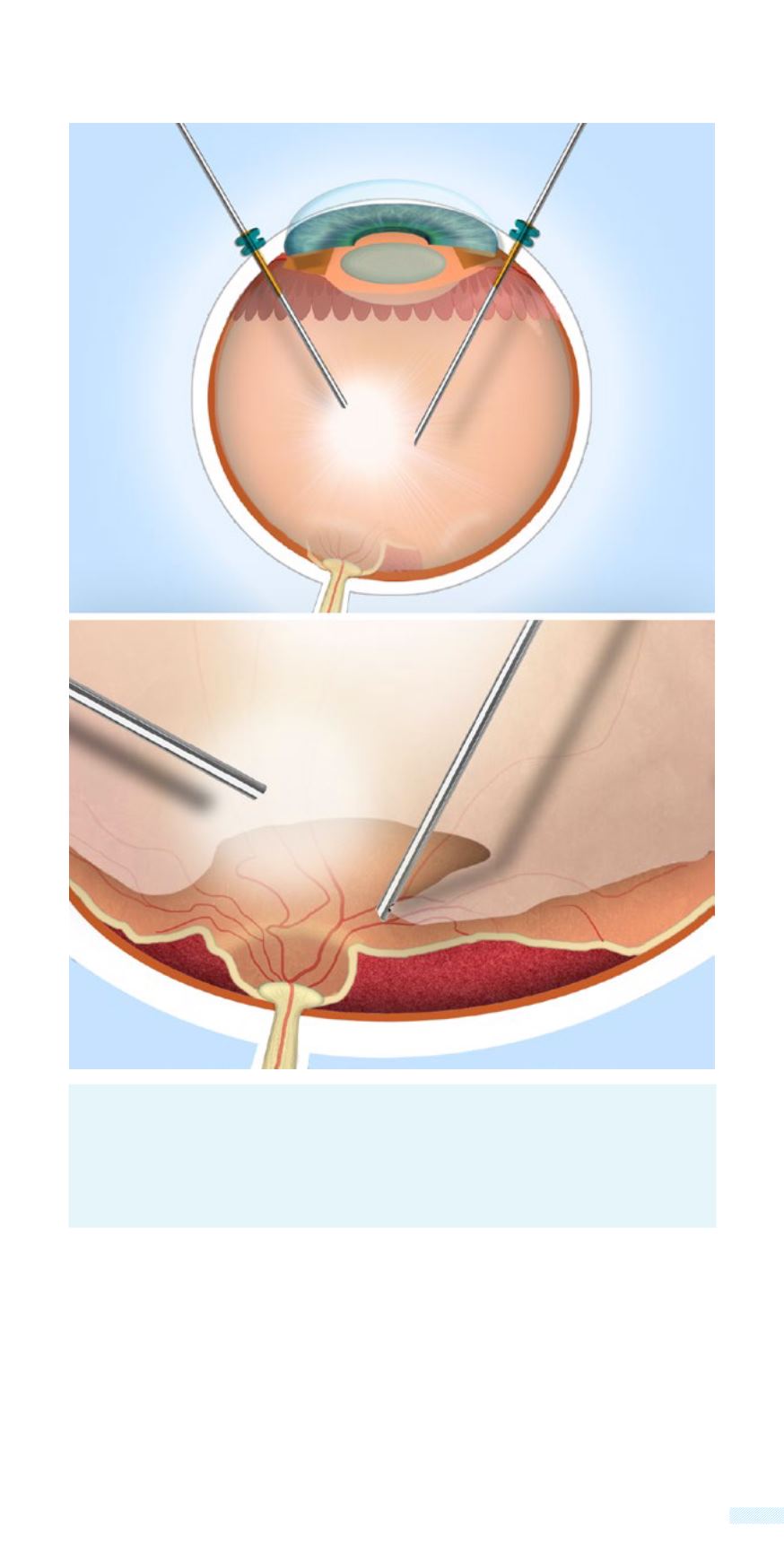
          Pars Plana Vitrectomy for Tractional Detachment.
        
        
          The tools for this
        
        
          operation enter the eye from an area called “the pars plana” from 3
        
        
          microscopical incisions, that remain open with the use of small trocars.
        
        
          All the manipulations for the separation of vitreous from the retina require
        
        
          great dexterity and experience, in order to avoid complications.
        
        
          ©
        
        
          2
        
        
          0
        
        
          1
        
        
          5
        
        
          N
        
        
          I
        
        
          K
        
        
          O
        
        
          L
        
        
          A
        
        
          O
        
        
          S
        
        
          P
        
        
          A
        
        
          P
        
        
          A
        
        
          Z
        
        
          O
        
        
          G
        
        
          L
        
        
          O
        
        
          U
        
        
          M
        
        
          .
        
        
          D
        
        
          .
        
        
          © 2015 ATHENS EYE HOSPITAL - NIKOLAOS PAPAZOGLOU M.D.
        
        
          Vitreotome
        
        
          Trocar
        
        
          Trocar
        
        
          Pars Plana
        
        
          Fibre light
        
        
          source
        
        
          Apart from the surgical technology,


            Athens Eye Hospital


          has at its
        
        
          disposal cutting edge technology for monitoring the surgery by
        
        
          the surgeon. The new ultramodern wide-angle system operating
        
        
          microscope  (OCCULUS BIOM) has a wide-angle field of view of 135
        
        
          degrees, and additionally it does not come into contact with the eye,
        
        
          but is adjusted on the surgical microscope, offering a panoramic
        
        
          view of the inside of the eye, with great sharpness and 3-dimentional
        
        
          viewing.