 German
 Ελληνικά
The retina is the very thin translucent membrane covering the inner surface of the eye. Its main role is to transform visual stimuli into nerve impulse, for analysis and processing by the brain.
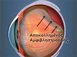 Under certain conditions the retina can detach from its position, resulting in devastating visual effects, if the detachment is not treated immediately.
The most common cause is the formation of a tear on the retinal surface, either spontaneously in predisposed patients (eg nearsighted), or after injury. Other conditions that can lead to detachment are ocular inflammation, vasculopathies, diabetes, etc.
Retinal detachment is a medical emergency and the patient should be able to recognize the symptoms of detachment in order to seek help as soon as possible.
These may include:
- Brief flashes similar to sparks, or lightning
- Sudden and mass appearance of moving black spots (floaters)
- A gray or black curtain that limits the visual field
- Blurred vision, that does not clear up with blinking or using artificial tears
These symptoms do not necessarily signify retinal detachment, but because the time is drawing in, the patient must urgently seek help from an ophthalmologist.
Treatment of tears that lead to detachment
Retinal tears usually only affect peripheral vision. Symptoms include unexplained flashes and the sudden appearance of numerous black spots.
The most common way of treating tears is to form a scar around the tear with a special Laser, which is done under local anesthesia. In doing so, the retina is repositioned and obstructed from further detachment, as well as the accumulation sub-retinal fluid, which can lead to detachment.
Scleral buckling
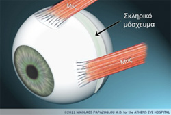 One of the most widespread ways to treat established retinal detachment is the scleral buckling surgery.
A silicon buckling element is sutured to the wall of the eye, in the area of the tear, applying inward pressure on the retina. This leads to the sealing of the tear and treatment of the detachment with the spontaneous reabsorption of the fluid collected under the retina.
The procedure is usually done under local anesthesia and the scleral buckles are permanently left in place.
Vitrectomy
In some types of detachment, such as tractional retinal detachment (fibrous tissue caused by inflammation or neovascularization pulling and detaching the retina), vitrectomy is chosen as the method of treatment.
During this procedure, the vitreous is removed (the gel normally found inside the eyeball). Then, the retina is flattened with the use of air or heavy liquid and is reattached with special endolaser.
Finally, the retina is stabilized by filling the cavity with a special gas (spontaneously absorbed in a few weeks) or, in more serious cases, silicone oil (which must be surgically removed after a few months).
A vitrectomy requires good post-operative cooperation from the patient, who must keep his/her head in a certain position (indicated by the doctor) for the next few days. This method, however, is less painful than the other methods and for many surgeons it is becoming the method of choice for retinal detachments.