 German
 Ελληνικά
The symptoms in the early stages of age-related macular degeneration may be subtle or nonexistent.
The surest way to diagnose eye diseases early on is to incorporate a complete eye examination in our annual check-up, especially after the age of 50.
Fundoscopy
An examination of the fundus (ie the interior of the eye) by an ophthalmologist is performed by using special lenses and can reveal the existence of age-related drusen (deposits) or other lesions in the macula.
Fluoroangiography and indocyanine green angiography
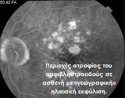 These are special techniques which photograph the fundus of the eye, which is preceded by the administration (injection into a vein in the arm) of a special dye. Areas of atrophy of the retina in a patient with "geographic" age-related degeneration.
These tests can give us valuable information about the condition of the vessels that runs into the eye, and have predictive value with concerns the a deterioration of central vision with scotomas or distortion of objects.
This distortion of objects (metamorphopsia) may mark the transition to the "wet form" of the disease and the presence of neovascularization and edema.
Although film has been replaced by digital cameras, a new innovation based on a special Laser has come to surpass even the digital technology.
SPECTRALIS ® HRA takes advantage of the precision of the special Laser and achieves even greater clarity and "depth" in its images, with the additional advantage that the 2 conventional angiographies (fluoro-and indocyanine) can be performed simultaneously, further reducing the patient's stay at the hospital.
Optical coherence tomography (OCT)
OCT is a diagnostic system that allows the analysis of all structures and layers in the macula. An OCT examination can reveal the most minute changes or damage in the retinal structure, it can evaluate and monitor the progress of diseases in the macular area, and the response to therapy.
Electrophysiology
Electroretinography (ERG) is a test which measures the "electricity" produced from the retina, where it receives a light stimulus. The region that normally gives the highest potentials, is the central fovea, the part of the retina that is most sensitive to stimuli. In the age-related macular these potentials are affected, and may even reach their complete suppression in advanced stages of disease.
Microperimetry
This is a test that is very similar to that of visual fields examination. The difference is that only the central region of the retina is examined and not the periphery.
With the assistance of the microperimetry, safe conclusions can be draw firm on the function of the macula lutea, whilst the same machine can be used to educate patients to make better use of the vision they have left.