 German
 Ελληνικά
Non-proliferative retinopathy
 At this stage the image of the fundus has microaneurysms, hemorrhages, exudates and edema in the retina. When leakage of components from the blood to the tissues occurs in the most central point of the retina (the macula lutea) then this is macular edema. The above is the result of the damage to the small vessels (capillaries) of the retina due to diabetes.
Proliferative retinopathy
Proliferative retinopathy
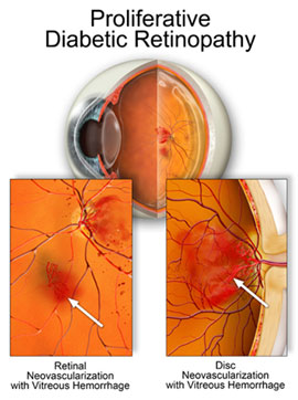 Due to poor blood circulation in the retina, low oxygen zones are formed (ischemia). In response to this condition, the eye creates new pathological blood vessels, which are very sensitive and can hemorrhage. The final stage of proliferative retinopathy includes hemorrhaging of the vitreous, scarring, detachment, neovascular glaucoma and vision loss.
Clinically significant macular edema
 Macular edema or swelling is more common in type II diabetes. Macular edema entails reduced or distorted vision.
Diabetic macular edema can be distinguished as:
- Focal, induced by microaneurysms and other vascular abnormalities that cause vascular leakage
- Diffuse, which concerns small capillaries that are located within the retina and cause diffuse swelling