 German
 Ελληνικά
It is a real shame for undiagnosed glaucoma damage to progress and deteriorate when there is diagnostic equipment available that can quickly and painlessly detect the slightest damage to the optic nerve.
HRT, GDX, OCT of the optic nerve
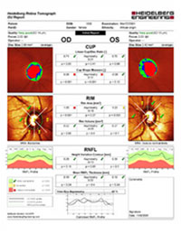 These acronyms represent three latest technology examinations that are designed for the purpose of detecting the slightest anatomical damage to the optic nerve, decisively contributing to the prevention of glaucoma.
All three tests are based on computer systems and take large international epidemiological studies into consideration in order to identify individuals who may potentially develop glaucoma.
Furthermore, in patients that already have glaucoma, the numerical recording of these measurements allow the monitoring of any deterioration over time, and the effectiveness of treatment.
Three-dimensional optic disc imaging
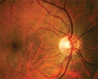 The head of the optic nerve, ie the entry point of the optic nerve to the eye, is called the "optic disc" and can be studied using special lenses.
The modern digital photography techniques provide accurate, extremely clear, three-dimensional imaging of the optic disc.
In glaucoma, even from its earliest stages, there are anatomical changes in the optic disc (thinning of the nerve fiber layers, increasing cupping, etc.) that can be assessed by a clinical ophthalmologist. It has a significant advantage compared to a simple fundoscopy given the ability of comparison with previous and subsequent examinations, thus ensuring an objective decision of its evolution over time.