 German
 Ελληνικά
The Vitroretinal department at the Athens Eye Hospital offers innovative technology to cover the full spectrum of manifestations of diabetic retinopathy with special investigations.
Topcon fundus camera (digital photography)
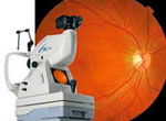 Color imaging of the fundus of the eye in order to record and display the patient's clinical picture on examination day.
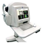
Repeat imaging over time helps in monitoring the development and various Cirrus Zeiss OCT detected retinal damages: There are 2 machines for faster patient service.
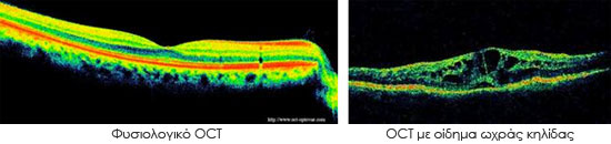
 The optical tomography (Optical Coherence Tomography) is the newest element used in the diagnosis of diabetic maculopathy, which is the major cause of reduced vision in diabetics.
The optical tomography (Optical Coherence Tomography) is the newest element used in the diagnosis of diabetic maculopathy, which is the major cause of reduced vision in diabetics.
Fluoroangiography and indocyanine green angiography
The Athens Eye Hospital has two different imaging machines for the angiography examinations. The Topcon system with Imagenet software and the Heidelberg spectralis (HRA).
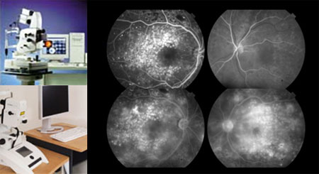
The test uses contrast dye (fluorescein or indocyanine) which is injected into a vein in the arm of the patient, which makes the blood vessels to glow and we can distinguish leakage, ischemia, abnormal blood vessels, swelling, etc. The fluoroangiography is a very important test in diabetic retinopathy, which allows monitoring of progression of the disease based on which we make important treatment decisions.
Ophthalmic Ultrasound (Ultrasound workstation Paradigm)
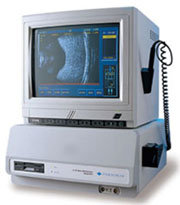 Ultrasound study of the eyes in diabetic patients with dense vitreous hemorrhage, or clouding of the transparent media, for diagnosis of retinal detachment and an abundance of other intraocular disorders that can coexist in a diabetic patient and especially in one with advanced damages.