 German
 Ελληνικά
Tonometry
This is the name given for measuring intraocular pressure. It is performed on each eye separately, and usually at different times on the same day to record a 24hour variation (tonometric curve).
The upper limit of normal values for intraocular pressure is considered to be 22 mmHg. In no case, however, is the pressure value to be considered a diagnosis, and must always be evaluated in combination all clinical and laboratory data to determine whether the patient needs treatment or not. Moreover, as previously mentioned, the values that are "within normal range" are not necessarily safe and do not exclude the possibility of an incipient or existing damage to the optic nerve.
Gonioscopy
Gonioscopy is the direct view of the angle by the ophthalmologist using a special lens that is applied to the cornea (ie touching the eye). This test can unambiguously determine the range of the drainage angle as well as any possible pathological deposits that can obstruct it.
There are several systems to grade the range of the angle, but the term "narrow angle" is usually used to describe the short "distance" between the iris and inner surface of the cornea which anatomically restricts the outflow of aqueous humor and may lead to an increase in pressure .
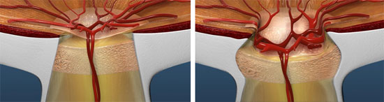
Fundoscopy examination of the optic disc
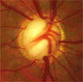 Even before glaucoma reveals its first symptoms, the ophthalmologist is able to determine the morphological changes of the head of a damaged optical nerve (which is called the "optic disc") with a simple fundoscopy examination
With this examination, which is ideally performed with the use of eye, the interior of the eye, which in its entirety is called the fundus, is examined via the eye's pupil (the black hole in the center of the colored iris).
The ophthalmologist will assess changes some of which are thinning of the nerve fiber layers, increased normal cupping the optic disc, altered the course of vascular route, microhemorages etc. in order to determine the extent of damage.
Perimetry (Visual fields)
The assessment of the extent of optic nerve damage, manifested by a reduction or loss of vision a part of the area normally from which the patient should normally see is with conducted using machines called perimeters or visual field analyzer or simply visual fields.
With this painless test, which takes several minutes for each eye, the patient is asked to confirm the lighting of a series of strategically placed light signals by pressing the button on a controller that he/she is given.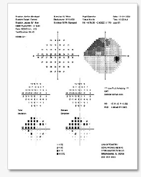
This is an examination that is absolutely necessary, since it shows the actual, functional visual impairment, as this perceived by the patient.
However, the visual fields examination begins to present findings when optic nerve damage is already extensive, established and irreversible.
Studies have shown that when the first damage appears in the visual field analysis, more than 30-40% of optic nerve fibers have already been lost . Thus, it is imperative to use new technologies in order for this damages to be identified as early as possible and avoid the bad consequences of glaucoma.